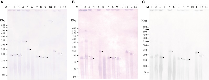
Figure 1.
Detection of plasmid(s) in the donors and transconjugants using Southern hybridization following to pulsed field gel electrophoresis with the tet(M) probe (A), traI-pAQU1 probe (B), and rep-pAQU1 probe (C). Lane: M, DNA size standard (lambda ladder); 1, 04Ya001; 2, 04Ya016; 3, 04Ya090; 4, 04Ya108; 5, 04Ya265; 6, 04Ya311; 7, TJ001W1; 8, TJ016W1; 9, TJ090W1; 10, TJ108W1; 11, TJ265W1; 12, TJ311W2 and 13, W3110. Approximately, 20 ng of DNA was loaded in each lane. Arrows indicate the positions of the bands.

