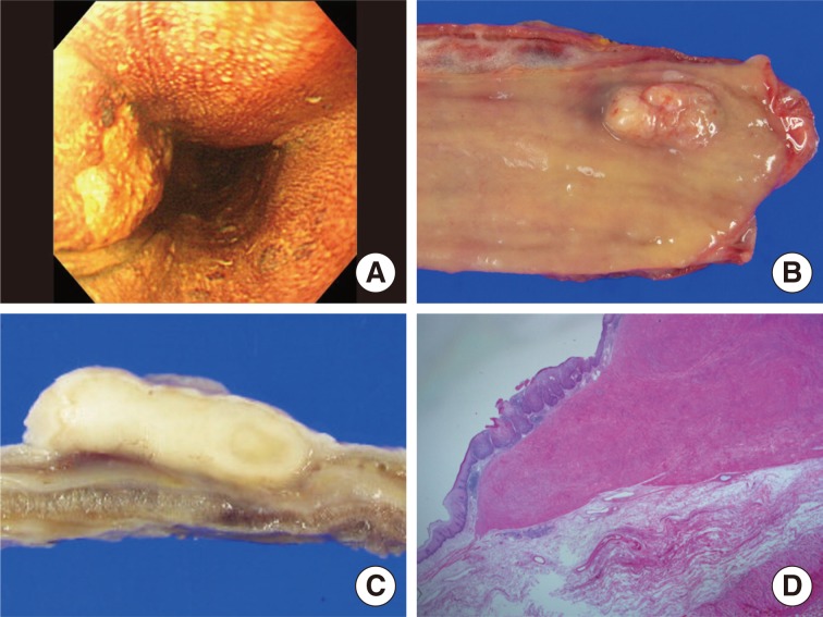
Fig. 1.
(A) Endoscopic examination reveals an esophageal tumor in the proximal third of the esophagus, clearly identified as a nonstaining area by Lugol's iodine solution. The esophagectomy specimen shows an elevated lesion with central ulcer (B) and the cut surface shows a well demarcated, white, solid mass (C). (D) The scanning view shows a well demarcated submucosal mass with an overlying abnormal epithelial lesion.

