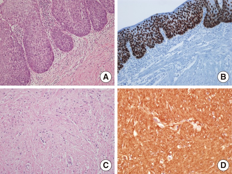
Fig. 2.
(A) Microscopic findings show squamous cell carcinoma in situ overlying submucosal leiomyoma. (B) The squamous cell carcinoma in situ component shows strong p53 immunopositivity. The submucosal spindle tumor is composed of interlacing bundles of bland spindle cells (C) and is immunoreactive for actin (D).

