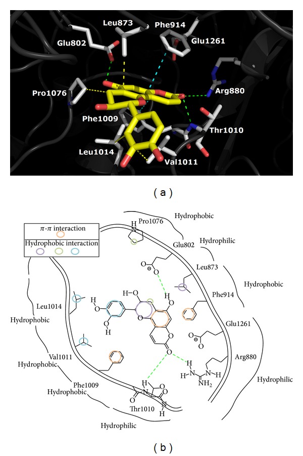
Figure 5.

Predicted binding mode of compound 5 docked into the active site of xanthine oxidase. The top and down pictures of each panel display the 3D and 2D structural docking patterns, respectively. The nitrogen and oxygen atoms are shown in dark blue and red colors, respectively. The hydrogen bond formation and the electrostatic interaction between compound 5 (yellow) and the amino acid residues (gray) of XOD are shown in green and light blue dashed lines, respectively.
