
Figure 2.
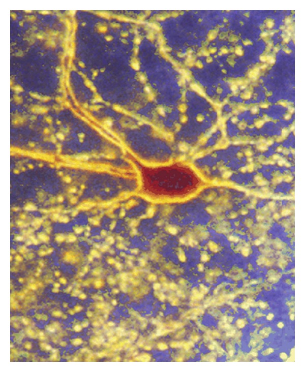
Photomicrograph of a cerebellar Golgi cell. The cell was juxtacellularly filled with neurobiotin and viewed in dark field at magnification of 240x. The soma, principal dendrites, and a diffuse cloud of axon terminals are visible, each terminal corresponding to a contact with one of thousands of granule cells (adapted from Holtzman et al. [73]).