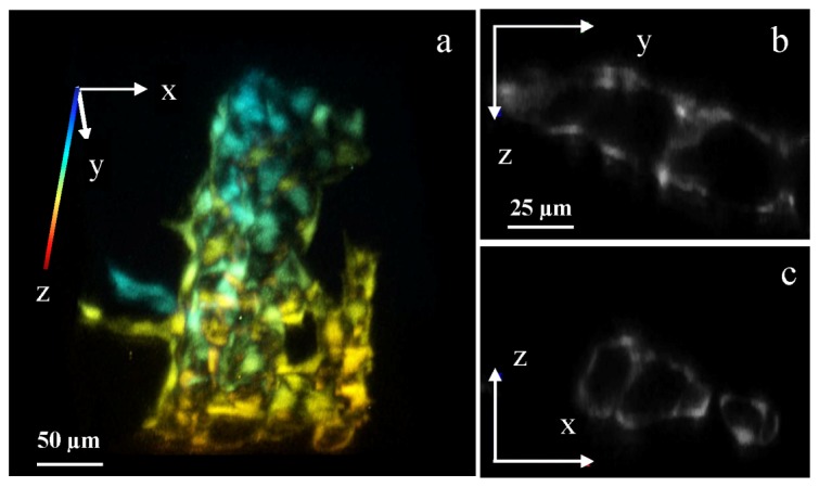
Fig. 4.

Cellular 3D imaging by two-photon Bessel light-sheet microscope. (a) 3D rendered image of epithelial cells forming vasculature in a Tg(kdrl:GFP) zebrafish tail. The 3D image is color-coded in z-axis. (b) Cross section in the y-z plane and (c) in the x-z plane, showing vessel structures. Full 3D image (512 × 512 × 88) can be viewed in Media 2 (7.8MB, MP4) . 3D images were acquired in 1-μm z-steps by moving the sample tube, with the imaging plane moving deeper into the fish as z increases. The tube lens focal length was set to 300 mm, resulting in a lateral pixel size of 0.27 μm. The exposure time was 1s per step.
