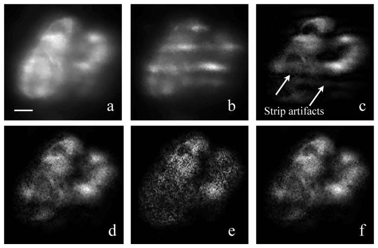
Fig. 9.

Diffusion reduction with a modified SIM reconstruction algorithm. (a) Merged image of 5 SIM raw frames with phase-shifted patterns. The merged image is equivalent to a 10-seconds-exposure image under uniform light-sheet. (b) Raw image frame under structured light-sheet illumination; the expose time was 2s; (c) Reconstructed image by the standard SIM algorithm, showing stripe-shaped artifacts due to residual diffusion background; (d) Reconstructed image from ± 1 order harmonics by the improved SIM algorithm. Stripe-shape artifacts were removed; (e) Reconstructed image from ± 2 order harmonics by the improved SIM algorithm; (f) Weighted merge of ± 1 and ± 2 order reconstructed image. All images were taken from the kidney of a live transgenic Tg(pod:NTR-mCherry) zebrafish at 4 dpf. Podocytes in the fish kidney is labeled with mCherry. Images was taken with the tube lens at f = 135mm and a lateral pixel size of 0.6 μm. The scale bar is 10 μm.
