Fig. 11.
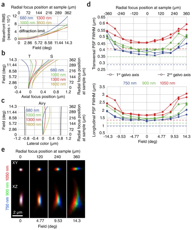
High-resolution color-corrected imaging with constant PSF to within less than 3.6% width variation over more than 400 µm Ø FOV. (a) Calculated wavefront error in the objective pupil for the complete laser scanning microscope from (Fig. 9) using custom scan and tube lenses (Fig. 6 and Fig. 8(b)) over 680-1600 nm and 720 µm FOV when using the Olympus XLPLN25xWMP objective. (b) Calculated field curvature for sagittal (S) and transversal (T) beam components, longitudinal color and astigmatism at the sample location for the complete laser-scanning microscope assuming an ideal objective. (c) Calculated lateral color at sample location assuming an ideal objective. (d) Measured transversal and longitudinal two-photon excitation PSFs along the two orthogonal directions of the scan mirrors over the whole FOV (continuous lines) of the complete two-photon laser scanning microscope using the Olympus XLPLN25xWMP objective and comparison to theoretical value (dashed lines) of an aberration-free PSF. (e) Lateral and longitudinal chromatic PSF aberration of individual microspheres measured at 750 nm, 900 nm and 1050 nm.

