
Fig. 3.
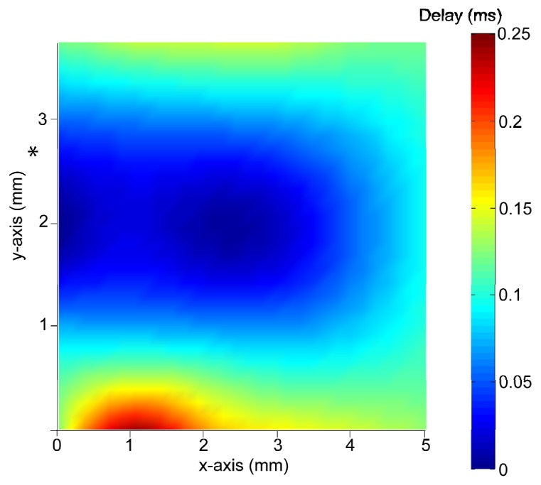
Elastic wave propagation speed by OCE in the rabbit cornea after CXL. Using a greater tissue stimulation force, it is possible to determine elastic wave propagation over the entire recording surface even in tissue that was made stiffer by the cross-linking treatment. Blue-shifted colors represent shorter time delay between measured points, corresponding to greater elastic wave propagation speed. (*) = point of tissue stimulation (n = 1).