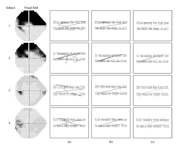
Figure 1.

Four examples of scanpaths from four different glaucoma patients with their visual fields on the left. The start and end of each saccade are represented by a circle. Column (a) shows the original scanpaths made by the four participants reading the text. Column (b) shows the scanpath after the rotation has been corrected and reading-specific saccades have been extracted using the preprocessing algorithm. Column (c) shows the scanpath results from the clustering and classification algorithm. The number represents the order in which the saccades occurred, and the colours represent the classification that was attributed to them by the automated clustering algorithm (blue: forward saccade, green: between line saccade, red: regression, and brown: unknown).
