Fig. 5.
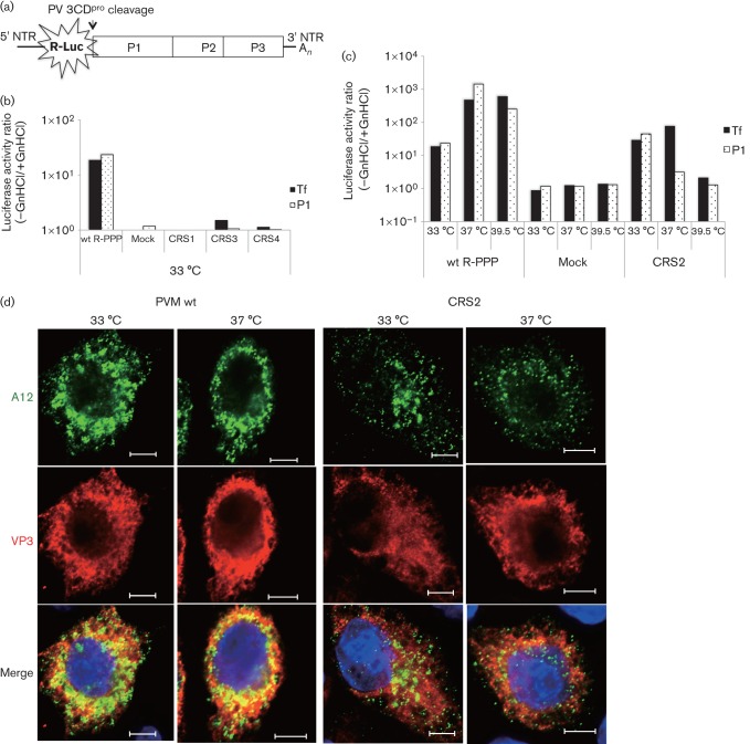
RNA replication and encapsidation phenotypes of the four CRS mutants. (a) The genome structure of the R-PPP virus is illustrated. R-Luc was fused to the N terminus of the polyprotein and cleaved off during translation at a 3CDpro cleavage site. (b) RNA transfections of wt and mutant RNAs were carried out in the absence (−) and presence (+) of GnHCl. Lysates were passaged onto fresh HeLa cells, in the absence and presence of the drug, and R-Luc activity was determined (see Methods). R-Luc activity produced by wt and mutant reporter viruses (CRS1, CRS3 and CRS4) after transfection (indicating replication) and after first passage (P1; indicating encapsidation) at 33 °C are shown. For the sake of simplicity, a ratio of R-Luc activity in the absence and presence of GnHCl is plotted on the figure. (c) Transfection and passage of the wt and ts CRS2 mutant were carried out at all three temperatures (33, 37 and 39.5 °C) and R-Luc activity was determined. Mock, no transcript RNA was added. (d) Immunofluorescence imaging of PVM wt and CRS2 virus-infected HeLa cells at 33 and 37 °C. HeLa cells were infected with wt or CRS2 viruses at an m.o.i. of 1 at 33 or at 37 °C. The infected cell cultures were incubated for 4 h (37 °C) or 5 h (33 °C). The infected cells were probed with primary antibody A12 (a gift from N. Altan-Bonnet, Laboratory of Host-Pathogen Dynamics, National Institutes of Health, Bethesda, Maryland, USA), which recognizes the mature virus, followed by Alex Fluor 488-conjugated secondary antibody (green). VP3 localization in the same cell was with a polyclonal antibody to VP3 followed by Alex Fluor 555-conjugated secondary antibody (red). The blue immunofluorescence is the DAPI staining of the nucleus. Bars, 5 µm.

