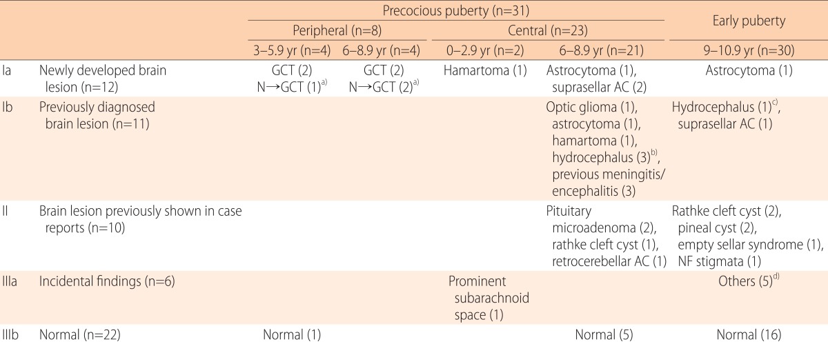
Table 1.
Brain magnetic resonance imaging findings in 61 boys with precocious or early puberty

GCT, germ cell tumor.
a)N→GCT: normal at first evaluation→newly develop germ cell tumor. b)Postoperative (n=2), intracranial hemorrhage (n=1). c)Postoperative (n=1). d)Prominent subarachnoid space(n=1), prominent perivascular space in basal ganglia(n=1), focal high intensity in white matter(n=1), cystic lesion in white matter(n=1), and cyst in coroidal fissure(n=1).
