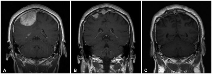
Fig. 2.
Enhanced coronal magnetic resonance imaging (MRI) series in a 45-year-old man (Case 11). A: Preoperative MRI showing a homogenously enhanced mass around right frontal convexity. B: MR imaging obtained 24 months after surgery and conventional radiotherapy revealing the recurrence of meningioma at previously surgical site with two separated enhancing mass. C: Thirty-seven months after operation with radiosurgery, a coronal MRI with gadolinium enhancement demonstrating disappearance of the meningioma.

