
Figure 2.
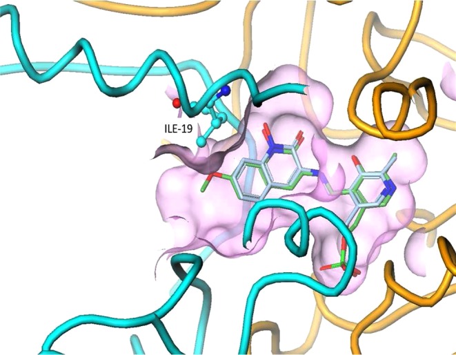
Superposition of the crystal structures of 1 and 2 with KAT II, showing the additional van der Waals interactions made by the methoxy substituent at C-7. The entire helix at the entrance of the binding pocket, consisting of residues Ile-19 to Gly-29, is disordered in the structure of PLP-bound 1 (yellow), whereas it is ordered in the structure of 2 (light blue).