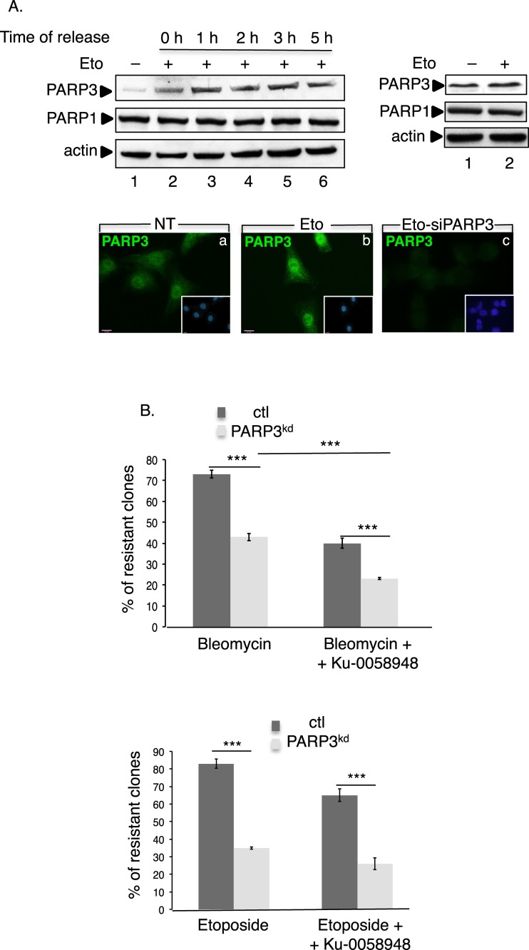
Figure 1.
PARP3 responds to and promotes survival upon the induction of double-strand breaks. (A) Enhanced expression of PARP3 upon etoposide treatment. Upper left panel: MDA-MB231 cells were either mock-treated (lane 1) or exposed to etoposide (Eto, 50 μM) (lanes 2–6) for 3 h and then released in fresh medium for the indicated time points (0 h, indicates no release). Equivalent amounts of nuclear extracts were analyzed by western blotting using the indicated antibodies. Upper right panel, MDA-MB231 cells were either mock-treated (lane 1) or treated with etoposide (Eto, 50 μM, 3 h, lane 2) and equivalent amounts of total protein extracts were analyzed by western blotting as above. Lower panel: representative immunofluorescence pictures showing the nuclear staining of PARP3 (green) in treated (b and c, Eto, 50 μM, 3 h) or nontreated (a, NT) MDA-MB231 cells. In (c), the etoposide treatment and immunofluorescence were performed 48 h after transfection with siPARP3. Insets: nuclei are stained in blue with DAPI. (B) The depletion of PARP3 sensitizes cells to DSB-inducing agents. Clonogenic survival of bleomycin-treated (upper panel) or etoposide-treated (lower panel) control (ctl) and PARP3kd cells in the absence or in the presence of the PARP1 inhibitor Ku-0058948 (100 nM). Experiments were performed >3 times giving similar results. Mean values of triplicates ±SD are indicated. ***P< 0.001. The depletion of PARP3 in the PARP3kd cells was verified by qRT-PCR (Supplementary Figure S1Ba).

