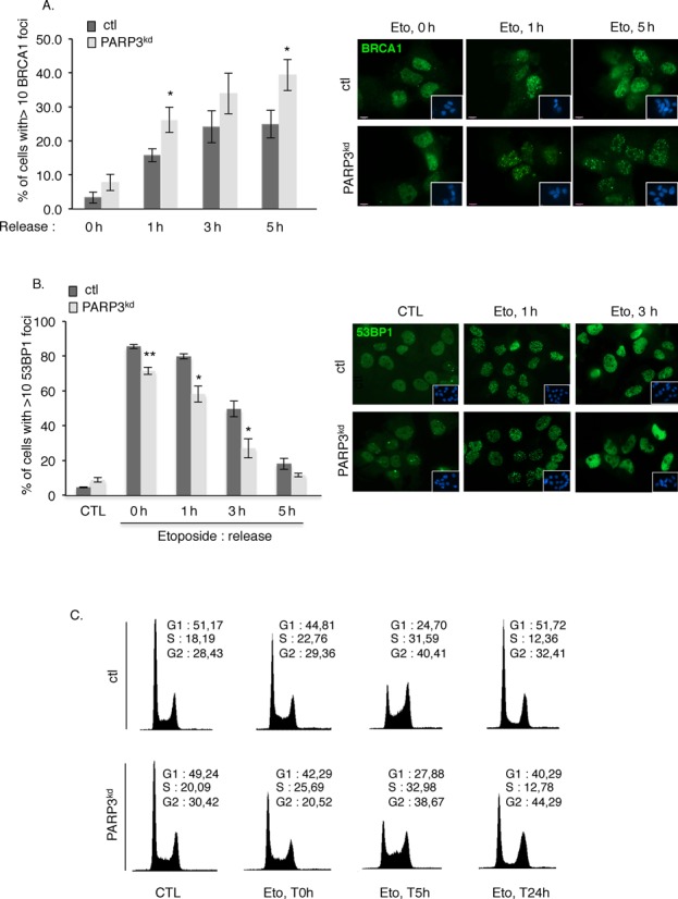
Figure 4.

PARP3 helps to modulate the balance between BRCA1 and 53BP1. (A) Increased accumulation of BRCA1 at DSB sites in PARP3-depleted cells. Control (ctl) and PARP3-depleted cells (PARP3kd) were treated with etoposide (50 μM) for 1 h, released in fresh medium and processed for immunofluorescence at the indicated time points using an anti-BRCA1 antibody. The histogram depicts the percentage of cells displaying >10 BRCA1 foci. An average of 500 cells per condition were scored in > 20 randomly selected fields. Data are represented as the means of three independent experiments ±SD. *P < 0.05. Representative immunofluorescences of the quantification of the positive cells with BRCA1 foci (green) are shown. Insets: nuclei are stained in blue with DAPI. A similar pattern of BRCA1 foci was observed in both cell lines. (B) Decreased loading of 53BP1 in PARP3-depleted cells. Control (ctl) and PARP3-depleted cells (PARP3kd) were treated with etoposide (50 μM) for 1 h, released in fresh medium and processed for immunofluorescence at the indicated time points using an anti-53BP1 antibody. The histogram depicts the percentage of cells displaying >10 53BP1 foci. An average of 500 cells per condition were scored in > 20 randomly selected fields. Data are represented as the means of three independent experiments ±SD. *P < 0.05; **P< 0.01. Representative immunofluorescences of the quantification of the positive cells with 53BP1 foci (green) are shown. Insets: nuclei are stained in blue with DAPI. A similar pattern of 53BP1 foci was observed in both cell lines. (C) PARP3-depleted cells display a similar S-phase delay as control cells after etoposide treatment but a sustained G2/M arrest. FACS analysis of ctl and PARP3kd cells mock-treated (CTL) or treated with etoposide for 3 h (Eto, 5 μM) and released in fresh medium for the indicated time points (0 h (non released), 5 h, 24 h). y axis: cell numbers; x axis: relative DNA content based on propidium iodide staining.
