
Figure 7.
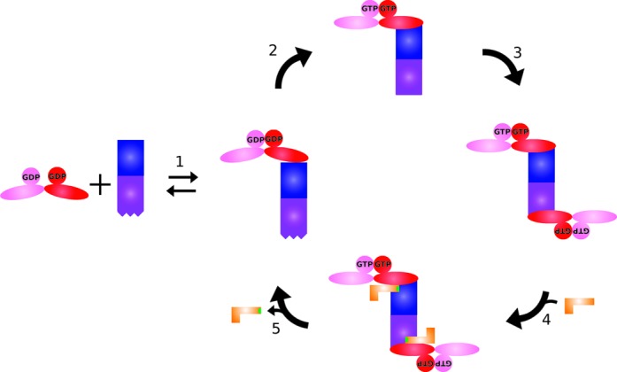
Model of MnmEG complex formation during the tRNA modification cycle (MnmE is colored red and pink; MnmG blue and purple). (1) MnmE is binding to MnmG in a non-symmetric manner with initial closure of the G domains. (2) Upon GTP binding the G domains of MnmE undergo further closing, with a concomitant movement of the helical domain, leading to a structural change in MnmG, (3) promoting the binding of a second MnmE dimer on the other MnmG subunit. (4) In the following step two tRNA molecules can bind mainly via interactions with MnmG and (5) upon GTP hydrolysis the tRNA becomes modified (indicated in green), the complex dissembles to its α2β2 form and the tRNA leaves the complex.