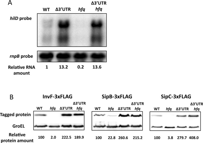
Figure 6.

(A) hilD mRNA levels in Hfq+hilD 3′UTR+, Hfq+hilD 3′UTR−, Hfq−hilD 3′UTR+ and Hfq−hilD 3′UTR− isogenic strains. hilD mRNA was detected by northern blot using a specific riboprobe, and rnpB mRNA was used as an internal control. For quantification, the ratio hilD mRNA/rnpB mRNA was relativized to 1 in Hfq+hilD 3′UTR+ background. (B) Western blots of InvF-3xFLAG, SipB-3xFLAG and SipC-3xFLAG in protein extracts from wild type, Δ3′UTR, Hfq− and Δ3′UTR Hfq− hosts. GroEL was used as loading control. For quantification, the ratio 3xFLAG-tagged protein/GroEL was relativized to 1 in the WT background.
