
Figure 2.
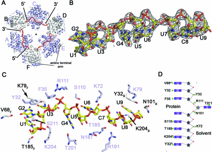
RNA binding to the N hexamer. (A) Surface representation of the Toscana virus RNP complex showing three RNA molecules in red bound to the inside of the hexameric ring. Binding of RNA induces a change from a six-fold to a three-fold symmetry (triangle). (B) 2Fo–Fc map contoured at 2σ showing electron density for the bound RNA oligomer. The asymmetric sequence allowed unambiguous fitting of the nonameric RNA into the electron density. (C) Cartoon representation showing selected contacts of N with the RNA. The RNA is capped on either side by Tyr32 of adjacent monomers. Residues depicted in white belong to the adjacent subunits (see Figure 2A for chain numbering). (D) A schematic diagram of the orientation of the RNA bases in the RNA-binding groove. RNA bases are recognized by both main and side-chain contacts.