
Figure 1.
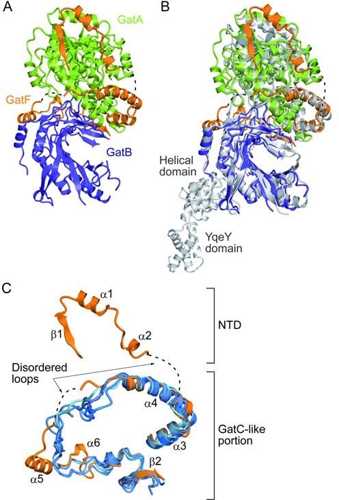
Structural comparison of GatFAB with GatCAB. (A) Ribbon representation of the crystal structure of S. cerevisiae GatFAB, consisting of the GatA (green), GatB (navy blue) and GatF (orange) subunits. Dashed lines indicate the disordered loops of GatF (residues 59–66 and 114–128). (B) The superposition of the crystal structure of S. cerevisiae GatFAB onto that of S. aureus GatCAB. GatFAB is shown in the same color code as in (A). S. aureus GatCAB is colored gray (PDB ID: 3IP4). (C) Structural comparison of S. cerevisiae GatF with the bacterial GatC. GatF is colored orange. The T. thermophilus, T. maritima, S. aureus and A. aeolicus GatCAB structures (PDB IDs: 3KFU, 3AL0, 3IP4 and 3H0R) are colored blue and superposed onto that of S. cerevisiae GatFAB.