
Figure 2.
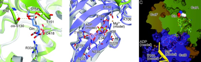
Active sites of GatFAB. GatFAB is colored as in Figure 1. (A) The glutaminase site of GatA in the Gln-bound form. The bound Gln molecule is shown as blue sticks. The crystal structure of A. aeolius GatA is colored gray and superposed onto that of S. cerevisiae GatA. (B) The amidotransferase site of GatB in the apo form. The crystal structure of S. aureus GatB is colored gray and superposed onto that of S. cerevisiae GatB. The modeled ADP molecule and Mg2+ ion are shown as pink sticks and as a sphere, respectively. (C) Surface model representation of a hydrophilic NH3 tunnel. The tunnel is filled with water molecules (red spheres), which interact with conserved residues of GatA and GatB. The modeled ADP and Mg2+ ion are shown as in (B). The modeled T. thermophilus tRNAAsn is colored yellow.