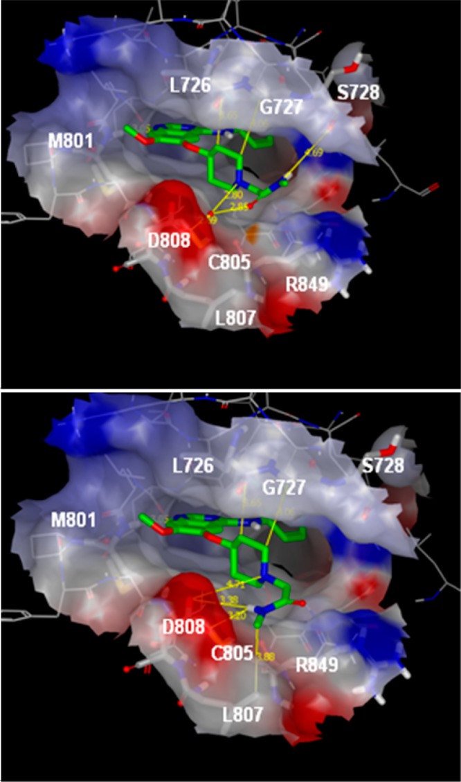
Figure 2.

Compound 2 positioned in the HER2 homology model based on the EGFR tyrosine kinase in complex with 4-anilino quinazoline inhibitor erlotinib (PDB code 1M17). The quinazoline N-1 binds to the hinge region at Met801, with the aniline buried deep in the selectivity pocket. The C6 alkoxy 4-piperidine sits at the solvent exposed rim of the ATP binding site making various hydrophobic contacts or CH···C=O short contacts with the surrounding residues (highlighted by yellow lines). The piperidine basic site is in the vicinity of the Asp808 residue offering an electrostatic complementarity with the cationic site. The molecule is shown in two representative conformations of the N-2-acetamide chain, conformation A (upper panel) and conformation B (lower panel). In the model corresponding to conformation A, a water molecule has been added to satisfy the interactions network. The protein surface is colored according to electronic surface potential. Areas in red indicate increasing negative potential, areas in blue increasing positive surface potential.
