Figure 6.
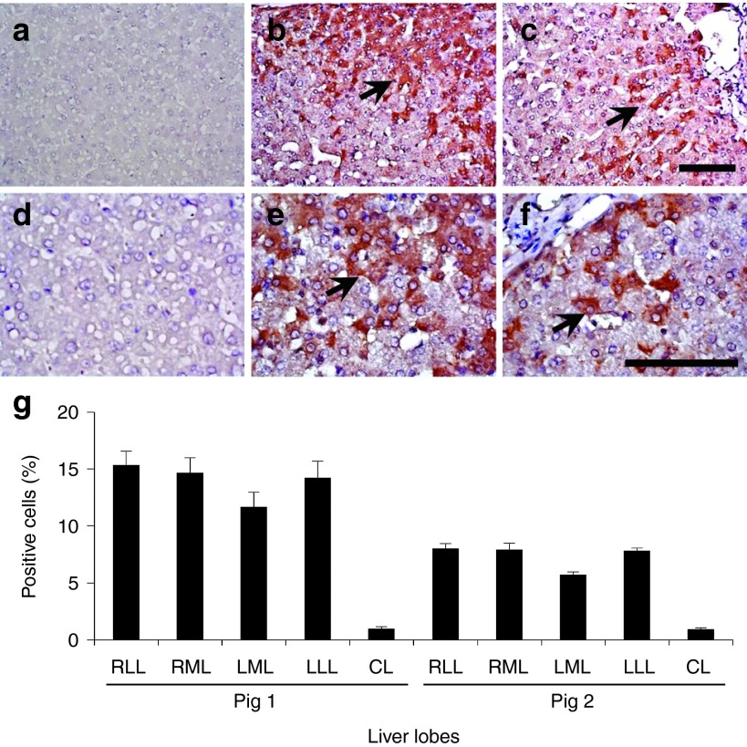
Persistent expression of human α-1 antitrypsin in the hepatocytes. Immunohistochemical staining of human α-1 antitrypsin was performed in the liver after the hydrodynamic gene delivery of pCAG-hAAT plasmid. (a, d) Noninjected CL in pig 1; (b, e) injected RML in pig 1; (c, f) injected RML in pig 2. Scale bar represents 100 µm (a, b, c, 200× d, e, f, 400×). Black arrows represent positively stained hepatocytes. (g) Quantitative analysis of positively stained cells. Ten liver tissue samples collected from each lobe (total of 50 samples in a liver) in pigs 1 and 2 and a quantitative analysis was performed on three fields (total of 150 fields in a liver) from each section. The values represent mean ± SD (n = 30 for each lobe). P < 0.05 between all injected lobes and CL. One-way ANOVA followed by Bonferroni's multiple comparison test.

