Figure 3.
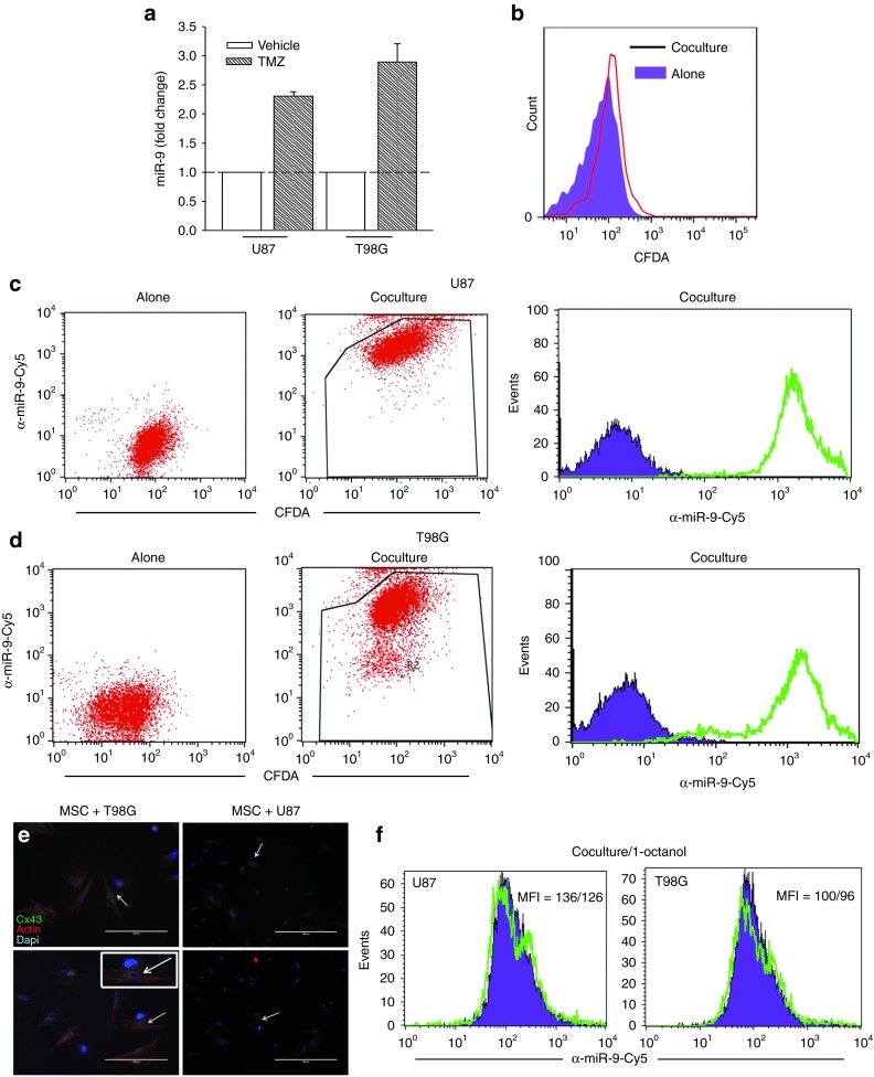
Transfer of anti-miR-9-Cy5 from mesenchymal stem cells (MSCs) to glioblastoma multiforme (GBM) cells. (a) Real-time polymerase chain reaction for miR-9 in U87 and T98G, untreated and treated with 200 µMol/l temozolomide (TMZ). The data for the TMZ-treated cells are presented as fold change in relation to vehicle, which is normalized to 1. (b) GBM cells were labeled with the cell tracker CMFDA and then placed in contact with MSCs. After 72 hours, the cells were analyzed for changes in Cy5. (c,d) Cocultures or transwell studies with anti-miR-9-Cy5-transfected MSCs and (c) CMFDA-loaded U87 or (d) T98G were studied for the transfer of Cy5 probe. Left panels show the GBM cells alone; middle panels analyze the GBM cells for CMFDA and Cy5; right panels show Cy5 alone in the GBM cells. (e) Representative immunocytochemistry is shown for Cx43 in cocultures of MSCs and U87 or T98G cells. The arrows show Cx43 (green) between two cells. Immediately before examining the cells, the cultures were labeled with phalloidin and diamidino-2-phenylindole. (f) Cocultures of U87 or T98G and MSCs were studied in the presence or absence of 300 µMol/l 1-octanol. After 72 hours, the cells were studied for Cy5.

