
Figure 2.
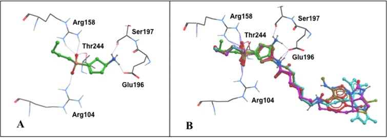
View of the ρ1 GABAC ligand-binding site with predicted binding modes. (A) Close-up of (S)-4-ACPBPA 6 (green carbons). (B) Close-up of (S)-4-ACPBPA-C5-NMA (S)-11d (brown carbons), (S)-4-ACPBPA-C5-NBD 12 (red carbons), (S)-4-ACPBPA-C5-BODIPY 13 (aqua blue carbons), and (S)-4-ACPBPA-C5-biotin 16 (magenta carbons) that are overlaid on (S)-4-ACPBPA 6 in the GABAC binding site, with the linker and fluorophores external to the orthosteric site.