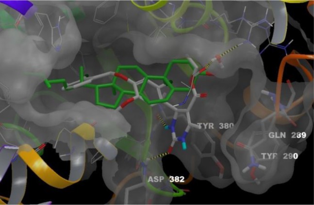
Figure 1.

Superposition of cholesterol binding mode with compound 8-(S) predicted binding mode. Ligands, cholesterol (green carbons) and 8-(S) (white carbons), are colored by atom type. The conserved water molecule appears as balls and sticks. Four residues from RORα binding site are labeled in stick representation: Gln289 and Tyr290 materialize the mobile loop; Tyr380 and Asp382 that interact with 8-(S) through two hydrogen bonds (yellow dashed lines). These bonds between the NH in N-3 position and the backbone of Tyr380 and between the C=O of the urea moiety and the backbone of Asp382 anchor 8-(S) in the RORα binding site. Residues 327–335 (front yellow helix) are hidden.
