
Figure 3.
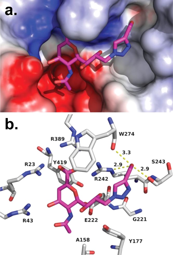
Molecular model of compound 6 in the active site of NEU4. Using our homology model of NEU4, compound 6 was docked to the active site and then subjected to molecular dynamics (10 ns). (a) An electrostatic surface representation of the active site is shown with compound 6 bound. (b) The general binding mode observed for DANA derivatives observed for NEU2 was preserved in our model, including contacts with the arginine triad (R23, R389, and R242). H-bond contacts are maintained between O4 and R43, as well as the glycerol side chain O8 with R242. The C4′–CH2OH has multiple H-bond contacts which include the backbone carbonyls of S243 (2.9 Å) and W274 (3.3 Å) and the Nε of R242 (2.9 Å).