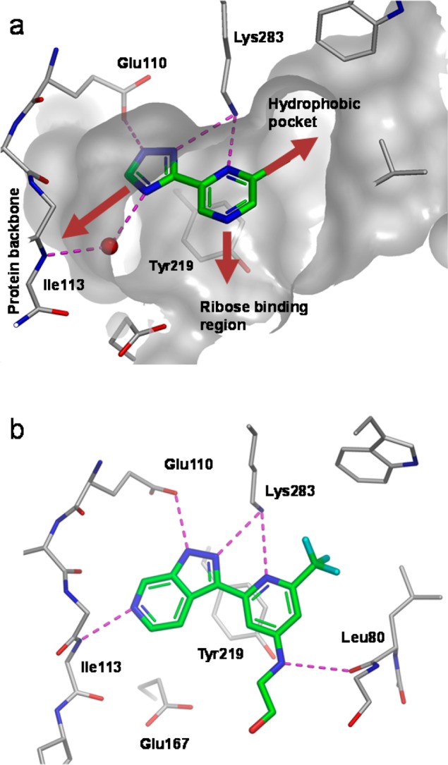
Figure 3.

(a) X-ray crystal structure of fragment 3 bound to LigA (S. aureus) showing key hydrogen bonds (purple dotted lines), a key water molecule (red sphere), and partially resected Connoly surface. (gray). Growth vectors toward the hydrophobic pocket, ribose binding region, and the protein backbone are shown by the red arrows. (b) X-ray crystal structure of 12 bound to LigA (S. aureus).
