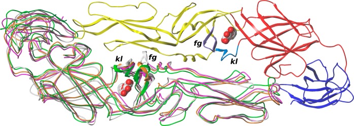
Figure 1.

Superposition of E protein structures and models for DENV (subunit 1 colored by domains (red, domain I; yellow, domain II; blue, domain III), subunit 2 colored magenta), TBEV (white), POWV (green), and OHFV (orange). Loops kl and fg are colored in subunit 1 and shown as ribbons in subunit 2. β-OG molecules are shown spacefilled.
