Figure 1.

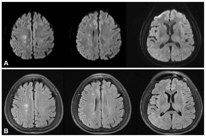
Diffusion-weighted (A) and fluid-attenuated inversion recovery (B) MR images demonstrate acute small infarctions in the right border zone between the anterior and middle cerebral artery.

Diffusion-weighted (A) and fluid-attenuated inversion recovery (B) MR images demonstrate acute small infarctions in the right border zone between the anterior and middle cerebral artery.