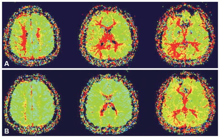
Figure 2.

Pre-stenting perfusion-weighted MR scans (A) show delayed mean transit on the subcortical area around the infarcted lesions. No definite evidence of hemodynamic insufficiency was found in the basal ganglia. Post-stenting follow-up perfusion images (B) demonstrate improvement of brain perfusion abnormalites in the right hemisphere.
