
Figure 5.
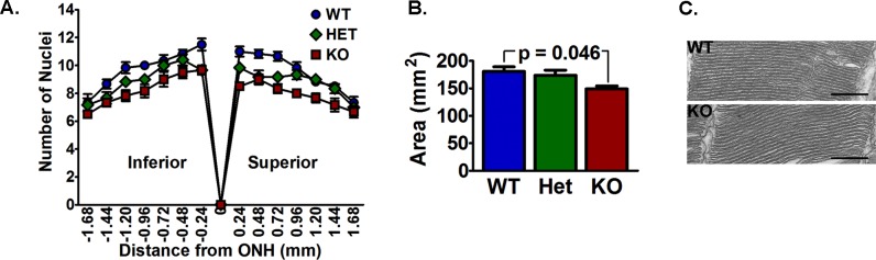
The VLC-PUFA–deficient mice had a loss of photoreceptors. (A) Histogram of the number of photoreceptor nuclei in a column at indicated distances from the optic nerve head to the peripheral retina. (B) Area occupied by photoreceptor nuclei measured from hematoxylin and eosin–stained sections was smaller in the KO mice than in the WT mice, but was not different compared to the Het retina (n = 5 WT, 7 Het, and 6 KO). (C) Representative micrographs of rod outer segments were similar between the WT and KO mice. Scale bars: 500 nm.