
. 2014 May 1;2014:140010. doi: 10.1530/EDM-14-0010
 This work is licensed under a
 Figure 1.
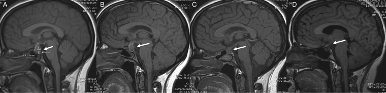
Cerebral MRI scans, sagittal T1. Arrows indicate the location of the mass. On admission and 1 week after, both pre-surgery (A and B): 1.7×1.1×1.9 cm rounded oval-shaped sellar lesion with suprasellar extension; immediately post-surgery (C) and 6 months post-surgery (D). We note the absence of the bright spot of the posterior pituitary on all four MRI scans. This can be predictive of a permanent central diabetes insipidus although specificity decreases with age.