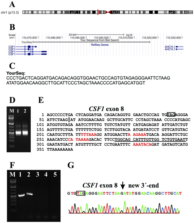
Figure 2.
Analysis of the new CSF1 transcript 5 in TSGCT. (A) Localization of the BLAT results on the chromosome 1 as it is obtained from genome browser. (B) Results of the BLAT search with one of the reads (YourSeq) which contained the last 20 nt of exon 8 of CSF1 (agtgtagagggaattctaag; nt 2081–2091 in sequence with accession no. NM_000757) and extracted from the raw sequencing data. (C) The sequence of the YourSeq which was used for BLAT search. (D) 3′-RACE on the cases 1 and 2 amplified a single cDNA fragment. (E) Sequence of the 3′-RACE-amplified cDNA fragment. The stop codon TAG is in box. The vertical line is the junction between exon 8 of CSF1 and the new sequence on 1p13 which is 41 kb downstream from the currently known CSF1 locus. The red letters are the polyadenylation signals. The primer CSF1-3end-R1out is underlined. (F) RT-PCR amplification using CSF1-1886F/CSF1-3 end-R1out primer combination. In case 1 (lane 1) and case 2 (lane 2), a single cDNA fragment is amplified. In case 3 (lane 3), the control cDNA (lane 4) and blank (no RNA in cDNA) (lane 5), no fragments are amplified. M is 1 kb Plus DNA ladder (GeneRuler, Fermentas). (G) Partial sequence chromatogram of the cDNA fragment amplified with primers CSF1-1886F/CSF1-3end-R1out showing the fusion of exon 8 of CSF1 with the new 3′-end.

