
Figure 3.
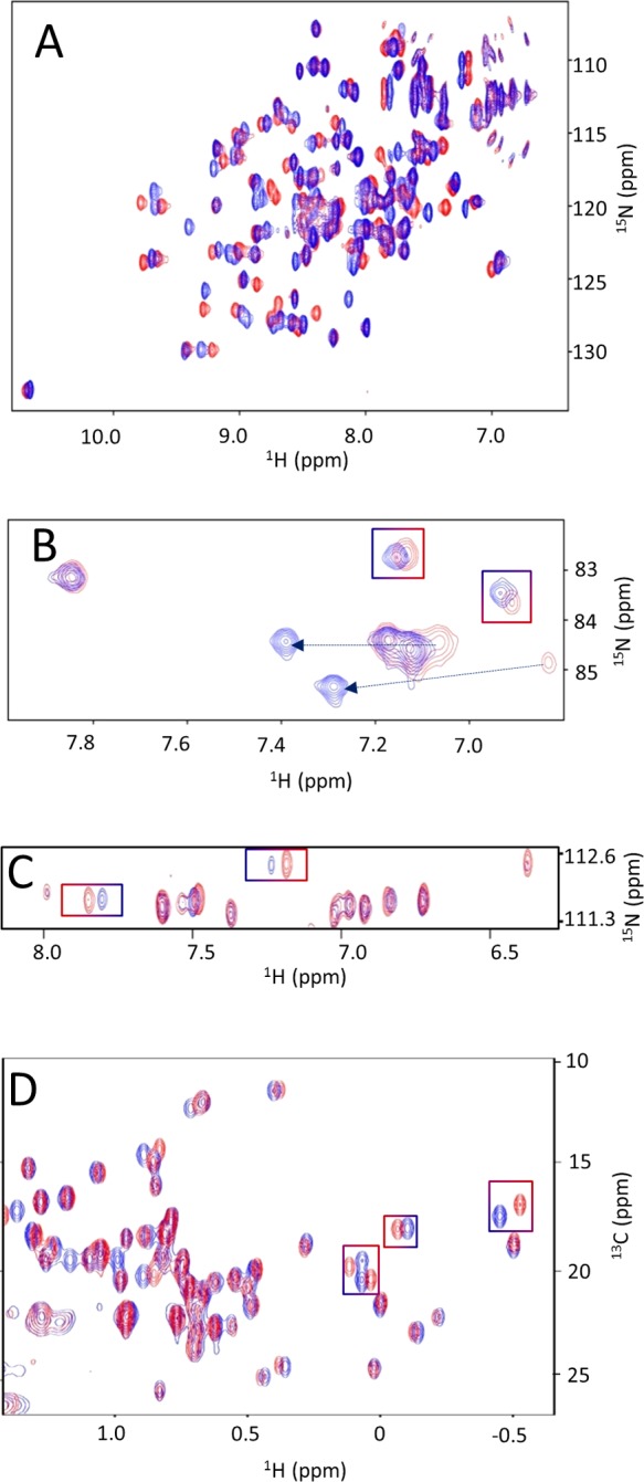
Examples of 2D protein-based NMR assays. Overlays of 2D heteronuclear NMR spectra of the vSrc-SH2 domain collected in the absence (red) and presence (blue) of a ligand. Panels A and B show [1H, 15N] SOFAST-HMQC spectra collected for backbone 1HN, 15N (A) or Arg 1Hε, 15Nε side chains (B). Panel C shows the spectra obtained with a modified [1H, 15N] HSQC (heteronuclear single-quantum coherence) experiment that selects for the 1Hδ, 15Nδ and 1Hε, 15Nε side chains of Asn and Gln, respectively. Panel D shows the [1H, 13C] HSQC spectra in the aliphatic region of the protein. Selected chemical shift perturbations in well-resolved regions of each spectrum are highlighted.