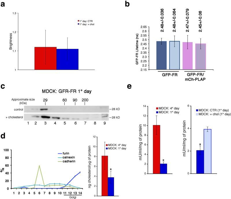
Figure 5. Cholesterol addition promotes homo-clustering of GPI-AP in the Golgi and regulates hetero-cluster formation and GPI-AP activity at the cell surface.
N&B (a) and FLIM (b) analysis were performed in 1-day MDCK cells pre-treated (2hrs) with cycloheximide in order to consider exclusively the cell surface pool of GFP-FR, in control conditions or upon cholesterol addition (in presence of cycloheximide). (c) 1 day MDCK cells treated with trypsin were subjected to temperature block, and incubated or not with cholesterol (during the last hour of temperature block) in order to analyse the GFP-FR Golgi pool and purified on velocity gradient. Molecular weight markers are indicated on top of the panels. (d) Cholesterol quantification after subcellular fractionation of MDCK cells. (Left) Quantification of the distribution of ER (calnexin), plasma membrane (cadherin), and trans-Golgi (furin) markers along the sucrose density gradient expressed as percentage of total proteins. (Right) Cholesterol amount in the Golgi-enriched fractions (11–14 fractions) quantified and normalized per microgram of protein in 4-days (red bar) and 1-day (blue bar) culture cells. Experiments were performed 2×. The error bars are the mean ± SD. *, p<0,005. (e) The alkaline phosphatase activity of PLAP in MDCK cells was measured (see Methods) upon different conditions: MDCK cells after 1 or 4 days in culture (left); 1-day MDCK cells in control or upon cholesterol addition (right). In all graphs the ALP activity of PLAP is expressed mU/ml and is normalized for total amount of protein. The error bars are the mean ± SD. *, p< 0,004.

