Figure 1.

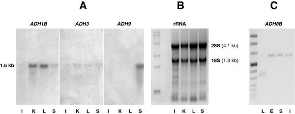
Detection of ADH classes in X. laevis. (A) Northern blot analysis of ADH1B, ADH3 and ADH9 from intestine (I), kidney (K), liver (L) and stomach (S), performed on 15-μg samples of total RNA. (B) Ethidium bromide-stained gel, from the same electrophoresis as in panel (A), showing 18S and 28S rRNAs next to the RNA molecular weight marker (0.24-9.5 kb, Invitrogen). The estimated molecular size of the RNA hybrids detected was ~1.6 kb. (C) RT-PCR of ADH8B from liver (L), esophagus (E), stomach (S) and intestine (I) next to DNA molecular weight marker VIII (Roche). Esophagus, stomach and intestine show an amplification product of 603 bp, indicating the presence of the ADH8B cDNA.
