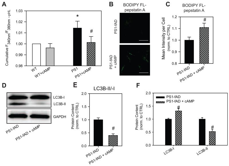
Figure 6. cAMP restores pHL and improves clearance in PS1-fAD fibroblasts.
A, Treatment of PS1-fAD cells with cAMP reduced lysosomal pH levels back to control levels. Treatment of control cells with the cocktail had a minimal effect. LysoSensor ratios normalized to mean control of each day’s experiment. N = 14 plates with 7-10 wells each. cAMP cocktail = 500μM cpt-cAMP + 100μM IBMX + 10μM forksolin. * = p <0.05 vs control, # = p <0.05 vs PS1-fAD alone. B, BODIPY FL-pepstatin A fluorescence is increased in PS1-fAD fibroblasts treated with cAMP cocktail. Scale bar = 100μM. C, Mean intensity per cell analysis of BODIPY FL-pepstatin A fluorescence reveals a significant increase in fluorescence; n = 60 PS1-fAD and 81 PS1-fAD cells. D, Western blot showing incubation with cAMP cocktail decreases LC3B-II/-I in PS1-fAD fibroblasts. E, Summary of 6 Western blots showing a significant decrease in LC3B-II/-I in PS1-fAD fibroblasts treated with the cAMP cocktail. F, LC3B-I is significantly increased in cAMP-treated fibroblasts, while LC3B-II is significantly reduced. For E, F, n = 11 PS1-fAD, 8 PS1-fAD + cAMP, 6 blots total. # = p < 0.05 for the entire figure.

