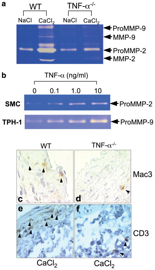
FIGURE 2.
Gelatin zymographic analysis of MMP-2 and MMP-9. a, Six weeks after NaCl or CaCl2 treatment, aortas from WT and TNF-α−/− mice were harvested. Aortic proteins were extracted and separated by electrophoresis on a 10% SDS-PAGE containing 0.8% gelatin. The gel shown is representative of three trials with similar results. The decrease in both MMP-2 (p = 0.003) and MMP-9 (p = 0.03) after CaCl2 treatment was significant. b, Up-regulation of the MMP-9 in macrophages and MMP-2 in mouse aortic SMC by TNF-α treatment. SMC and THP-1 cells, a human macrophage cell line, were treated with TNF-α for 24 h at concentrations of 0, 0.1, 1.0, and 10 ng/ml. Conditioned medium was separated on a 10% SDS-PAGE containing 0.8% gelatin for determination of MMP content. The gels are representative of four experiments with similar results. c and d, Immunohistochemistry of macrophage infiltration in the aortas of WT and TNF-α−/− mice after CaCl2 induction. Aortas were collected from WT and TNF-α−/− mice at 6 wk after CaCl2 treatment. Paraffin sections were immunostained with Mac3 Ab. Arrows indicate macrophage infiltration. Each staining represents three to four samples of WT or TNF-α−/− mice with similar results. e and f, Immunohistochemistry of T cell infiltration in the aortas of WT and TNF-α−/− mice after CaCl2 induction. Aortas were collected from WT and TNF-α−/− mice at 6 wk after CaCl2 treatment. Paraffin sections were immunostained with CD3 Ab. Arrows indicate T cell recruitments. Each staining represents three samples of WT or TNF-α−/− mice with similar results.

