
Figure 1.
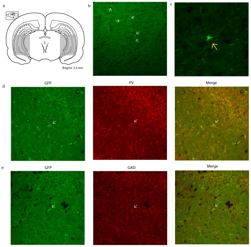
GFP+ MGE-derived cell transplants migrate throughout the hippocampus and adjacent regions. (a) Schematic depicting the approximate span of transplanted cell migration. (b) A representative hippocampal section displaying transplanted GFP+ MGE-derived cells (arrows) at a minimum of 2 months post-transplantation (10× magnification). The cell represented by a yellow arrow is displayed at a higher magnification (40×) in (c). Representative GFP+ MGE-derived transplanted cells stained for PV and GAD were imaged at 25× and are depicted in (d & e). Approximately 45.7% of the transplanted cells are positive for GAD, while 18.1% are PV positive.