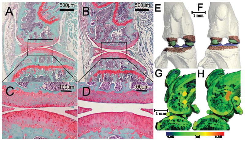
Fig. 1.
Histological images of a 184 day old wild type (A and C) and 182 day old integrin α1-null (B and D) medial condyle, stained with hematoxylin, fast green, and safranin-O (C and D are magnifications of the boxed areas in A and B respectively). Note that proteoglycan staining intensity, cartilage thickness and surface integrity and the size and abundance of chondrocytes are independent of genotype. Frontal (E and F) and sagittal through the medial condyle (G and H) microCT images of the same wild type (E and G) and integrin α1-null (F and H) knee as shown in A and B respectively. Trabecular bone is shaded red, subchondral bone blue, and ossified menisci green in E and F; cortical bone at the femoral condyles and tibial plateau was rendered transparent to show the analyzed trabecular regions. Sagittal sections (G and H) shaded with a thickness map with thicker regions shaded with colors located further right on the spectrum shown below. Trabecular bone architecture, subchondral bone thickness, and the size of ossified menisci and sesamoid bones are similar between the integrin α1-null and wild type knees.

