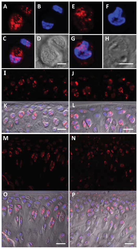
Fig. 6.
Basal phosphorylated Smad2/3 (pSmad2/3) in wild type and integrin α1-null chondrocytes. Wild type (A–D) and integrin α1-null (E–H) chondrocytes or sections of femoral condyle (I–L) and tibial plateau (M–P) chondrocytes from wild type (I, K, M, O) and integrin α1-null (J, L, N, P) mice stained with anti-pSmad2/3 antibodies (red) (A, E, I, J, M, N), Hoescht 33342 (blue) (B, F), overlay of both (C, G), DIC channel (D, H) and DIC channel overlaid on fluorescence (K, L, O, P). Scale bar = 5 μm (A – H) or 20 μm (I – P).

