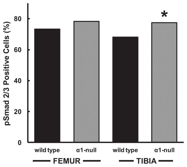
Fig. 7.
Graph of percent of basal pSmad2/3 stained chondrocytes in the femoral or tibial cartilage from wild type or integrin α1-null mice. A larger proportion of integrin α1-null chondrocytes (α1-null) stained positively for pSmad2/3 compared to wild type controls, reaching statistical significance in the tibia. ‘*’ indicates significantly (P<0.001) different from corresponding wild type control. Data from a total of at least 244 femoral (n≥244) and 433 tibial chondrocytes (n≥433) from N=3 mice and n≥14 sections were graded in each group.

