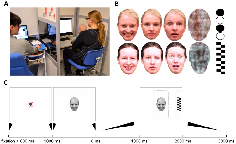
Figure 1. Eye tracking setup and the paradigm.
A) The photograph shows the whole study setup where infant is placed into a baby carrier attached to the chest of the caregiver (right), and the experimenter (left) is sitting behind a light wall with one-way transparency (not visible on the photograph) to allow observation of the infant during test protocol. One-way transparency was created by tinting the window and having higher lighting level on baby’s side. B) The two sets of face stimuli used in our study. Every infant was presented stimuli from one set only (chosen randomly). Both sets included a face with happy, neutral, and fearful expressions, as well as a noise (or ‘non-face’) stimulus that was created by a phase-scrambling of a face image to retain many physical image properties. Peripheral distractors (called ‘target’) consisted of checkerboards and other geometrically simple high-contrast stimuli. C) Content of a trial. Each trial was preceded by showing a fixation cue in the center of the screen to attract infant’s attention. The trial started only after infant’s fixation in the central area (small square around the dot) had lasted 600 ms. Then, the face stimulus was shown for 1000 ms, followed by adding the target to either side of the screen for the remaining 3000 ms. The stippled lines depict areas of interest (AOI) used for computing MD and DT metrics. The AOIs were deliberately defined to be larger than the original stimuli to cancel out effects of measurement inaccuracies and to reduce unnecessary noise in DR traces (see also figure 2 and Discussion on spatial accuracy in Supplementary data).

