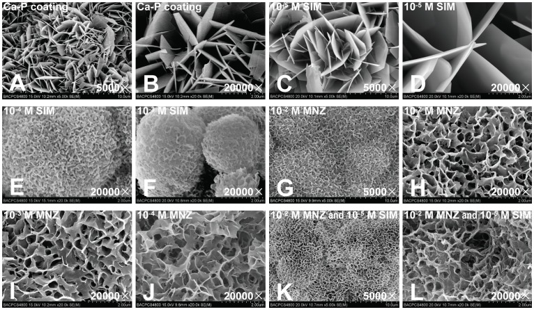
Figure 1. Scanning electron microscopy (SEM) observations of the Ca-P coating and drug-loaded Ca-P coating.
(A, B) Ca-P coating. (C, D) Ca-P coating loaded with 10−5 M SIM. (E, F) Ca-P coating loaded with 10−4 M and 10−3 M SIM. (G, H) Ca-P coating loaded with 10−2 M MNZ. (I, J) Ca-P coating loaded with 10−3 M MNZ and 10−4 M MNZ. (K, L) Ca-P coating loaded with 10−2 M MNZ and 10−5 SIM together.

