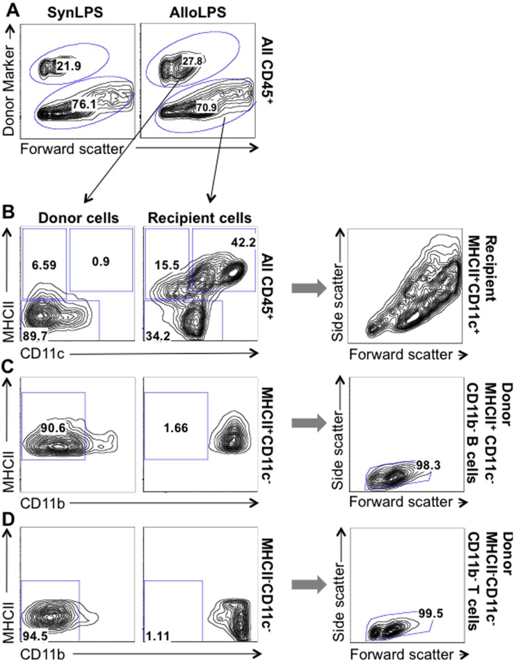
Figure 3. After allogeneic lymphocyte transfer followed by inhaled LPS, pulmonary donor-derived cells are comprised primarily of lymphocytes while myeloid cells are of recipient origin.
Rag1−/− mice received a transfer of allogeneic (Allo) or syngeneic (Syn) splenocytes and underwent daily exposures to aerosolized LPS for 5 days starting 1 week after splenocyte transfer. Mice were euthanized 72 hours after the last LPS exposure and lung cells were analyzed using flow cytometry. For flow analysis, a singlet gate was used to exclude cell aggregates, followed by an all-cell gate to exclude small debris and dead cells, followed by a CD45+ cell gate to define all white blood cells (as described in methods). (A) All CD45+ cells were separated into donor and recipient-derived cells based on their expression, or lack thereof, of the donor marker CD45.1 (in the case of SynLPS mice on the left) or H2Kk (in the case of AlloLPS mice on the right). The subsequent graphs show flow cytometry plots for AlloLPS lung cells but the same analysis was performed for SynLPS and yielded similar results. (B) Donor- (left) and recipient-derived (right) lung cells were analyzed separately. Representative flow cytometry plots show gating of MHC+CD11c+ antigen-presenting myeloid cells, which are mainly present in the recipient cells (42.2% vs. 0.9% in donor cells) and are large based on the side by forward scatter graph (far right). The MHCII+CD11c− cells are classically comprised of B cells and CD11b+MHCII+ myeloid cells. MHCII−CD11c− cells are usually comprised of neutrophils, monocytes, and T cells. (C) Representative flow cytometry plots show the population of MHCII+CD11c− cells and gating of the CD11b− B cells that are more abundant among donor cells compared to recipient cells (90.6% vs. 1.66%). The far right graph, showing a side by forward scatter plot, demonstrates that these donor MHCII+CD11c−CD11b− B cells are indeed small cells. (D) Representative flow cytometry plots show the population of MHCII−CD11c− cells and gating of the CD11b− T cells that are more abundant among donor cells compared to recipient cells (94.5% vs. 1.11%). The far right graph, showing a side by forward scatter plot, again demonstrates that these donor MHCII−CD11c−CD11b− T cells are indeed small cells.

