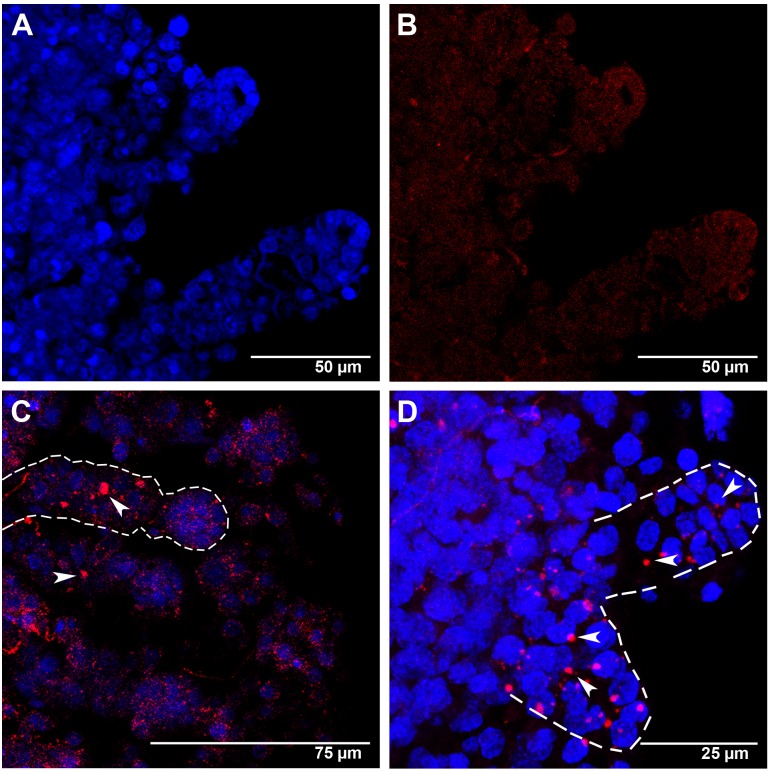
Figure 4. Amark transcript localization in worker ovaries at the L5F phase of the fifth larval instar.
(A) Ovarioles showing DAPI-stained nuclei. (B) The same ovarioles as in A, but labeled with the AlexaFluor555-Amark sense probe (FISH negative control), shows only a reddish background coloration. (C and D) Ovarioles labeled with the AlexaFluor555-Amark antisense probe and DAPI: the dashed line in C highlights an ovariole with large Amark foci (red) in the intermediary region (arrowheads). Amark foci (arrowheads in D) are also concentrated at the apical region of some ovarioles (shown in higher magnification and outlined by dashed lines in D).

