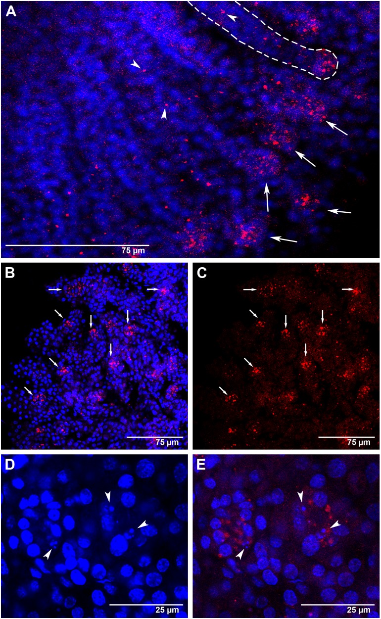
Figure 5. Amark transcript localization in worker ovaries at the L5S and PP phases of the fifth larval instar.
FISH with AlexaFluor555-labeled Amark antisense probe (red foci). Cell nuclei stained with DAPI (blue). (A) An L5S-phase ovary showing Amark transcripts highly concentrated at the apical end of the ovarioles (arrows). Amark foci are also seen outside the apical region (arrowheads). This pattern of Amark labeling is generalized throughout the worker ovaries. (B and C) At the end of the fifth larval instar (PP phase) the ovary continues to show Amark transcripts concentrated at the apical end of the ovarioles (arrows). (D) Detail showing small-sized degenerating nuclei (arrowheads) at the tip of the ovarioles (PP phase). (E) The same ovary as seen in D, but showing Amark foci (arrowheads) in the region where degenerating nuclei were identified.

