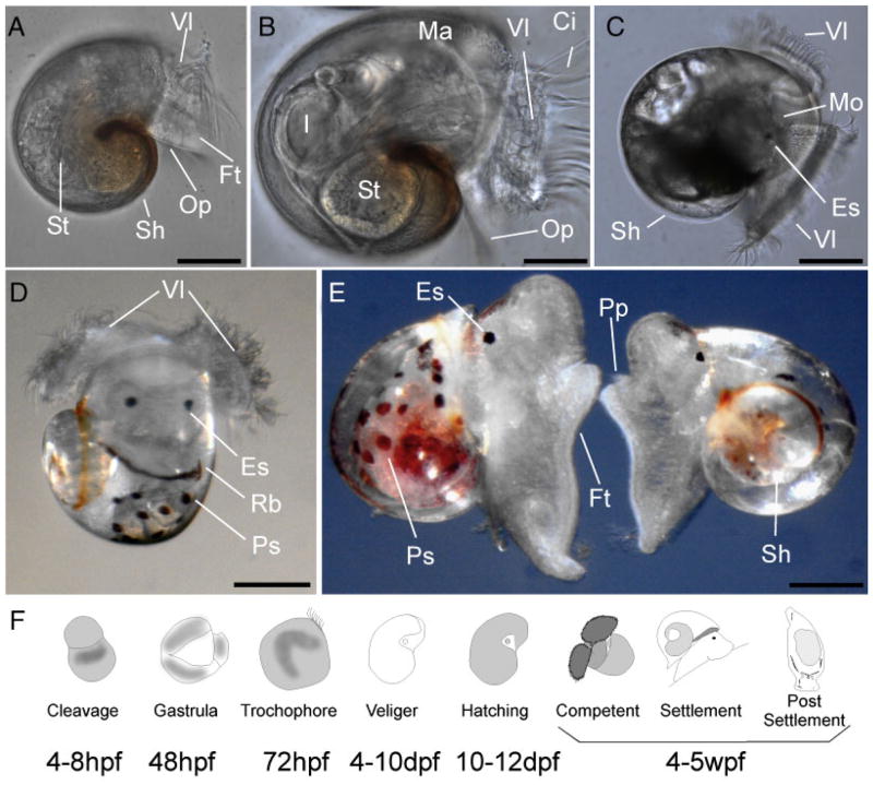
Figure 2.
Posthatching development and metamorphosis of A. californica. After hatching, the veliger larva will develop over an extended period of time (up to 3 weeks) in the plankton until all adult organs and the complete adult nervous system are formed (A–E; see Fig. 3). Only small changes in external morphology are visible during this time, but major changes occur internally, such as the development of the nervous system (see Fig. 3), the heart, and the excretory system. At the end of the planktonic period, the Aplysia veliger larva will reach metamorphic competence (D). Morphological characters for this stage are the occurrence of pigmented spots (Fig. 2D; Ps) and a red band that is visible on the mantle margin (Rb). Within 12–24 hr, competent larvae will transform into a settled juvenile (E; see Fig. 3). We are using the symbols indicated in panel F to describe the following stages: (I) early cleavage, (II) gastrulation, (III) trochophore, (IV) first veliger, (V) hatching, (VI) metamorphic competence (stage 6; see text for details), (VII) postmetamorphosis (stage 7; see text for details), and (VIII) 60 hr postmetamorphosis. The numbers below the icons indicate the approximate time line of embryonic and larvae development, based on our own observations at 20°C. Ci, cilia; Es, eyespot; I, intestine; Ma, mantle region; Mo, mouth; Op, operculum; Pp, propodium; Ps, pigment spot; Sh, shell; Vl, velar lobe; St, stomach. Panel E modified from (Heyland and Moroz, 2006). Scale bars: A: 45 μm; B: 50 μm; C: 90 μm; D: 90 μm; and E: 90 μm.

