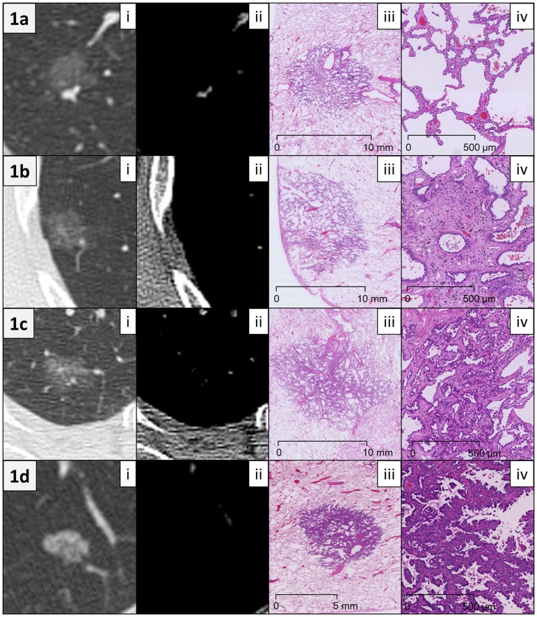
Figure 1. Representative radiological and histological images.
(a) A 64-year-old female patient with adenocarcinoma in situ. (i) Computed tomography (CT) scan (lung window setting) showed a pure ground-glass nodule (GGN), 11.9 mm in size. The mean CT attenuation of the tumor was −716 Hounsfield units (HU). (ii) Mediastinal window setting CT showed no tumor components except for vessels. (iii) Low magnification image (hematoxylin and eosin (HE) staining) showed a circumscribed tumor growing purely with a lepidic pattern without foci of invasion. A slight thickening of the alveolar walls in the tumor area was observed. (iv) Middle magnification image of the tumor (HE staining) revealed that tumor cells appeared to replace normal pneumocytes on alveolar walls. (b) A 63-year-old female patient with minimally invasive adenocarcinoma. (i) Lung window CT image showed a pure GGN, 14.2 mm in size. The mean CT attenuation was −691 HU. (ii) Mediastinal window CT showed no tumor components. (iii) Low magnification image of the tumor (HE staining) revealed a subpleural tumor consisting predominantly of lepidic growth with a small (<5 mm) focus of invasion. (iv) Middle magnification image of the invasive area of the tumor (HE staining) revealed acinar-type growth pattern. (c) A 74-year-old female patient with lepidic-predominant invasive adenocarcinoma. (i) Lung window CT showed a pure GGN, 19.7 mm in size. The mean CT attenuation was −618 HU. (ii) Mediastinal window CT showed no tumor components except for vessels. (iii) Low magnification image (HE staining) revealed a tumor consisting mostly of lepidic growth with a smaller area (8 mm) of acinar invasion. (iv) Middle magnification image of the invasion area of the tumor (HE staining) revealed acinar gland proliferation in the fibrous stroma. (d) A 76-year-old male patient with papillary-predominant invasive adenocarcinoma. (i) Lung window CT image showed a pure GGN, 10.7 mm in size. The mean CT attenuation was −509 HU. (ii) Mediastinal window CT showed no tumor components. (iii) Low magnification image of the tumor (HE staining) revealed that the tumor predominantly consisted of papillary proliferation. (iv) Middle magnification image of the tumor (HE staining) revealed cuboidal tumor cells growing along fibrovascular cores in a papillary configuration.

